
Niederlassung SLV München


Scanning electron microscope
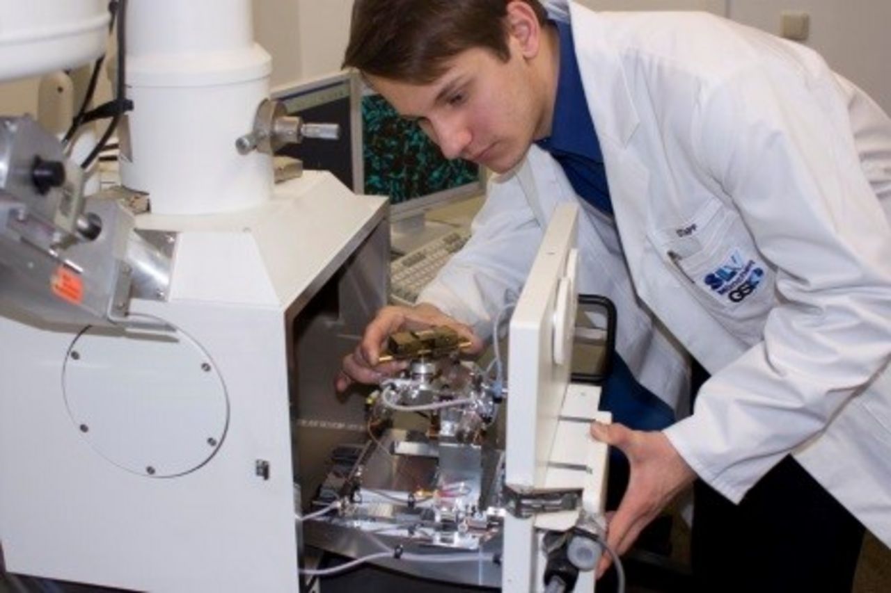
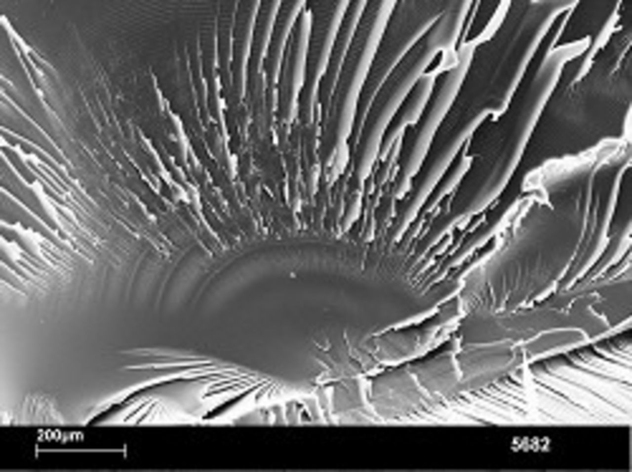
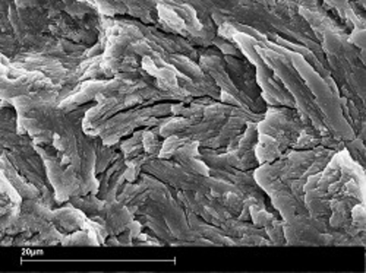
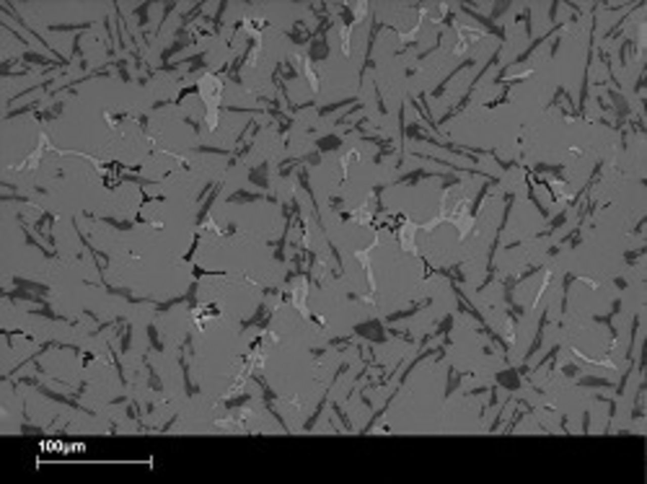
With the scanning electron microscope factographic examinations of fractures or in the laboratory opened crack areas are made as part of damage analyses.
These examinations are essential for the clarification of cases of damage. Because of the high depth of field of the scanning electron microscope different fracture structures, fracture origins, fracture and crack spreading directions can be represented as well as in the material formed foreign phases, excretions and corrosion coverings on the fractured surface.
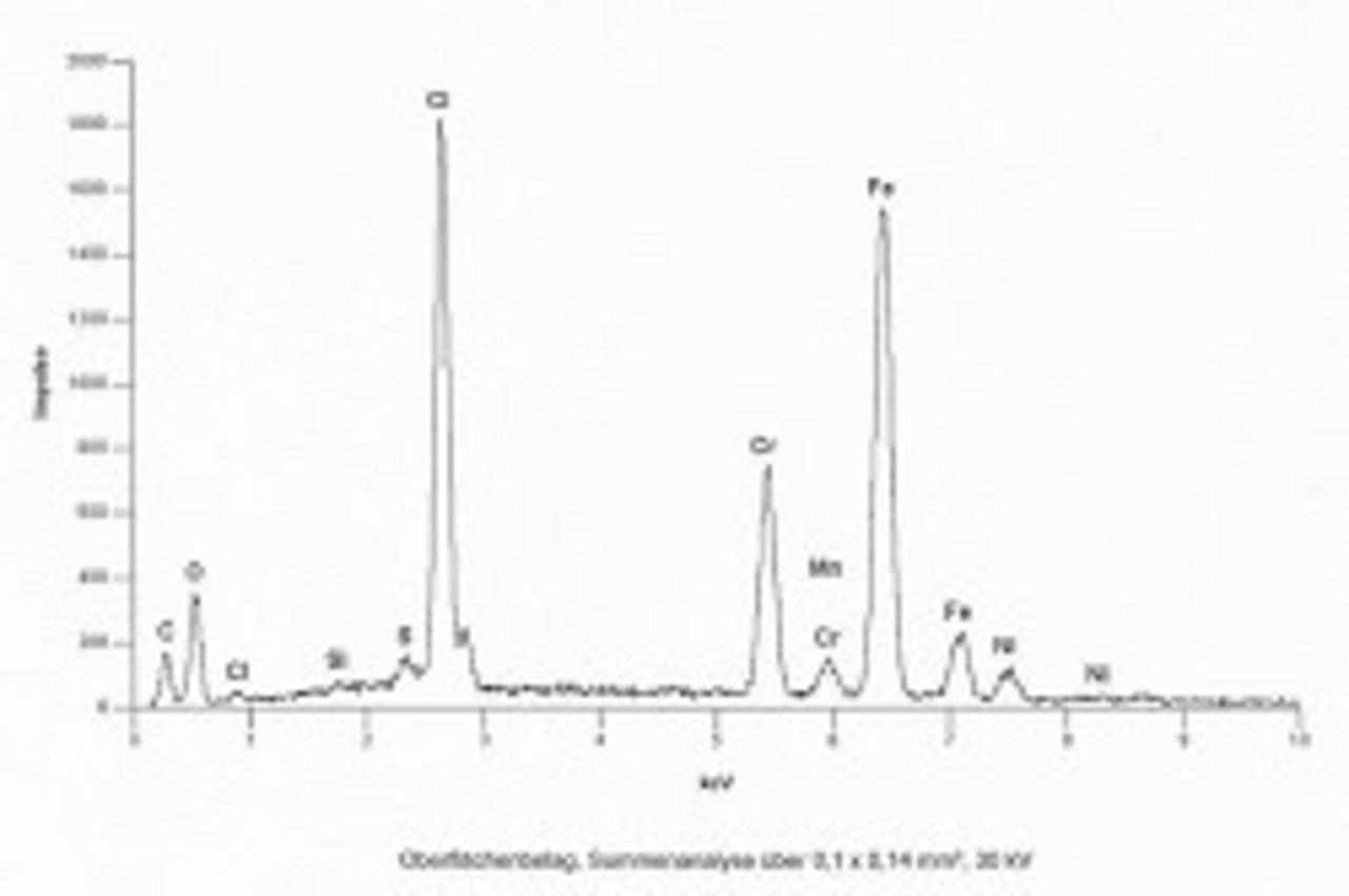
Electro beam microanalysis respectively energy dispersive x-ray analysis
On the basis of electro beam microanalysis (EMA/ESMA), also called energy dispersive x-ray analysis (EDX) the quality of the existing elements can be proven within minutes - independent of the state of the surface. in the direct EMA- image smallest parts can be located and identified with a spot analysis.
The EMA enables for example the determination of the chemical composition of corrosion products and can detect the finest metallographic constituents and excretions, whereby conclusions to the damage triggering factors, material irregularities or material specialities can be made.
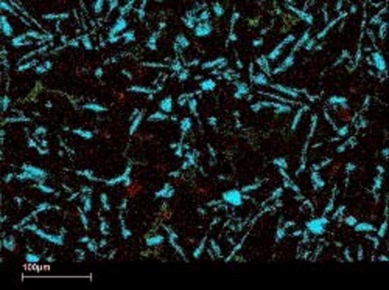
Element distribution - Mapping
At the scanning electron microscope element distribition images can be created with the help of EMA. Thereby within a previously defined area the concetration changes of the element distribution can be detected and simultaneously shown colored for 3 elements.